Osteoscopy:
Exploration of an osteoporotic bone using a mini-camera
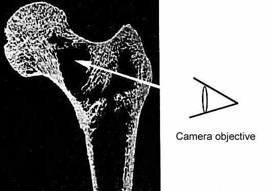
The camera penetrates the bone through a
12 mm hole, made with a bone file on the outer surface of
the greater trochanter. In this area the wall of the bone
is very thin. In the same way as holes made in the abdominal
wall for the passage of various instruments are closed with
sutures or staples, the hole in the bone is closed by refitting
the bone removed to allow the camera through.
Just as we work with flowing water in the knee, so the interior
of the bone is irrigated to remove any particles it contains
which are engendered by the osteoporotic disease. These are
essentially abnormally mobile cells and fat. These gradually
replace the bone structure. The more advanced the osteoporosis,
the larger the number of these particles. As a consequence,
the camera will be able to make progress more easily.
:: Close ::
|